
ASD Closure Surgery

UNDERSTANDING
ASD CLOSURE SURGERY
Minimally Invasive & Open Heart
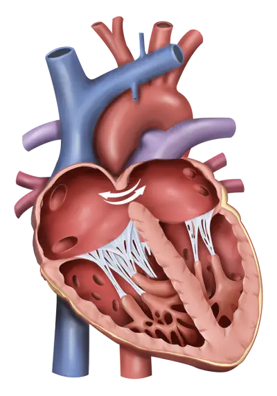
Some people are born with congenital heart defects, such as an atrial septal defect (ASD). This condition involves a hole between the heart’s two atria, which can cause shifts in heart pressure and potentially lead to strokes. When indicated, individuals, often young, may seek keyhole closure of this defect. This minimally invasive procedure, in experienced hands, offers rapid relief and a quick return to normal activities.
APPROACHES
Minimally Invasive
An endoscope, a flexible tube with a camera and light, is inserted through one of the incisions to provide a clear view of the heart and the atrial septal defect.
A catheter is threaded through a vein, typically from the groin, and guided to the heart and the site of the defect.
A closure device is delivered through the catheter. The device is positioned to cover and close the hole in the atrial septum.
The position and effectiveness of the device are verified using echocardiography (ultrasound of the heart) and sometimes fluoroscopy (X-ray imaging).
Open Heart
Almost all ASDs can be easily and safely closed through a small incision on the chest.
CONDITIONS TREATED
Surgery is recommended for patients with:
Minimally Invasive
- ASDs
Open Heart
- ASDs
WHAT TO EXPECT
Minimally Invasive
- Thorough assessment including medical history, physical examination, and advanced imaging (e.g., echocardiogram, CT scan).
- Consultation with (Adj) Professor Theo Kofidis to explore minimally invasive & open heart options, their potential benefits & risks, and if patient qualifies for minimally invasive surgery.
Open Heart
- Thorough assessment including medical history, physical examination, and advanced imaging (e.g., echocardiogram, CT scan).
- Consultation with (Adj) Professor Theo Kofidis to explore minimally invasive & open heart options, their potential benefits & risks, and if patient qualifies for minimally invasive surgery.
Minimally Invasive
- General anaesthesia is administered.
- A small incision is made in the femoral vein (and sometimes also the femoral artery) in the groin.
- A catheter holding the closure device is threaded through the vein to the heart.
- The device is placed into the hole, and the catheter is removed.
Open Heart
- General anaesthesia is administered.
- An incision is made in the chest to access the heart.
- The heart-lung machine is used to take over the work of the heart.
- The defect is located using an endoscope and closed with a patch, plug, or sutures.
Minimally Invasive
- Usually 1 day in ICU & 3-4 days in general ward.
- Faster mobilisation and return to daily activities.
- Follow-up appointments to monitor healing progress and overall cardiac health.
Open Heart
- Usually 2 days in ICU & 5-7 days in general ward.
- Slower mobilisation while in recovery.
- Follow-up appointments to monitor healing progress and overall cardiac health.
BENEFITS
Minimally Invasive
Less pain & quicker recovery period.
Results in smaller scars that are less visible.
Regain strength and mobility faster, allowing you to resume normal activities sooner.
Smaller incisions reduce the risk of post-surgical infections, leading to safer recovery.
Minimises blood loss, decreasing the need for transfusions.
Reduced physical trauma to the body facilitates quicker healing and less post-operative pain.
Lowers the likelihood of irregular heartbeats, enhancing overall cardiac stability post-surgery.
Preserving chest structure promotes improved lung function and breathing capacity shortly after surgery.
Smaller incisions result in less visible scarring, maintaining a more natural appearance.
Open Heart
In cases where the heart anatomy is complex or the arteries are severely diseased, open heart surgery may offer a more reliable option for complete revascularization.